Saturday, December 16, 2006

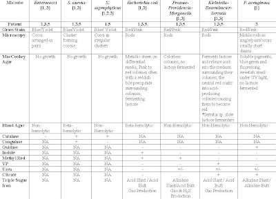
Compilation for Patients Diagnosed with UTI

Table 1: Suspected Organisms, Relevant tests and expected results of tests for respective patients diagnose with UTI

ABBREVIATIONS
NA - Not applicable


REFERENCES
1) Geo FB, Janet SB & Stephen AM. (2004). Jawetz, Melnick, & Adelberg’s Medical Microbiology. 23rd edition. McGraw-Hill.
2) Kenneth Todar. (2004). Staphylococcus. On site http://www.textbookofbacteriology.net > Google Search > Retrieved on 5th December 2006.
3) Wikipedia. (2006). On-site: http://en.wikipedia.org > Search > Retrieved on 4th December 2006.
*Note: So Sorry for posting late as we occurred some problems while posting

Thursday, December 14, 2006

SUSPECTED MICROORGANISMS (COMPILED)


The above are the suspected microorganisms for all five patients.

Thursday, December 7, 2006

MAYBELLINE WRITES!

Patient’s Name: Khong Fay Fay
Age: 26 Years
Sex: Female


Clinical Diagnosis
Complaints: Fever, chills, dysuria (pain during urination)
Diagnosis: Urinary Tract Infection
Antibiotic treatment: Nil

Urinary Tract Infection (UTI)
UTI is an infection that can happen anywhere along the urinary tract. It is usually caused by a bacterium from the anus entering the urethra and then the bladder. UTI is more common in women because their urethra is shorter and closer to the anus. (1) It may also be caused by dehydration, obstruction, disturbance of smooth urinary flow or presence of a foreign body, e.g. stone or urinary catheter. Trauma during sexual intercourse may also cause and infection in women. (2)

Usually, an UTI infected patient will experience dysuria, hematuria (bloody urine), fever, cystitis ( pain in the midline suprapubic region and/or frequent urination) and urethritis (discomfort or pain at the urethral or a burning sensation) .

The more common and suspected microorganisms that cause UTI are Escherichia coli (E.coli), Klebsiella pneumoniae, Proteus mirabilis, Psedonmonas aeruginosa. Staphylococcus saprophyticus and Enterococcus spp.

Gram-negative E.coli, Klebsiella and Proteus
E.coli, Klebsiella and Proteus are the most common cause of UTI. These bacteria fall in the family of the Enterobacteriaceae. The Enterobacteriaceae are a large, heterogeneous group of gram-negative rods. They are the most common group of gram-negative rods cultured in the clinical laboratory and the most common bacteria that cause disease. They are facultative anaerobes and are either motile with peritrichous flagella or non motile; they grow on peptone or meat media; grow well on MacConkey’s agar; ferment rather than oxidize glucose, often with gas production; are catalase-positive, oxidase-negative, and reduce nitrate to nitrite. (3)

Gram-negative Pseudomonas aeruginosa
Pseudomonas are commonly found in soil and water. P. aeruginosa sometimes colonizes humans and is the major human pathogen of the group. It is widely distributed in nature and is most found in moist environments in hospitals. P. aeruginosa tend to cause disease in humans with abnormal host defenses. They are gram-negative, motile and aerobic rods that are oxidase-positive and do not ferment lactose. (4)

Gram-positive Staphylococcus saprophyticus
10-20% of sexually active, young women are infected by Staphylococcus saprophyticus causing UTI. Staph. saprophyticus are gram-positive, cocci in irregular clusters and are facultative anaerobes. They are coagulase-negative and catalse-postive. (5, 6)

Gram-positive Enterococcus spp.
Entercococcus spp. are classified under Group D of the heterogeneous group of the Streptococci family. Streptococci are gram-positive spherical bacteria that form pairs or chains during growth. Group D Entercocci are usually non-hemolytics and occasionally alpha-hemolytic and are PYR positive. (7)

Diagnostics Laboratory Tests
The diagnosis of UTI can be confirmed by a urine culture. The urine culture will be sub cultured, isolated to identify the bacteria by running a series of appropriate biochemical tests. An antibiotic susceptibility testing will also be done to see which antibiotic is most suitable to be used to treat the patient. In diagnosis of UTI, if the urine culture yielding a greater than 100,000 colony-forming units (CFU)/mL and pure growth would indicate a significant bacteria growth. Or a urine culture that yield >100,000 CFU/mL with 2 bacteria growth may also indicate significant growth. However, if more than 3 organism is present, it indicates mixed bacteria growth and may be due to bad sample collection or contamination.

The urine culture is sub-cultured into TSA with 5% sheep blood agar (blood agar) and Cysteine Lactose Electrolyte (CLED agar).
The agar plates are left to incubate in the incubator at 37o C for 18-24hrs. The media is streak in a systematic manner as shown below.


After incubation, a gram stain would be done by isolating a colony obtained from the blood agar. A gram stain is done and examined microscopically to determine if the microorganism is a gram-negative or gram-positive bacterium. On microscopic examination, a purple-blue indicates gram-positive organisms while red-pink indicates gram-negative organisms.

GRAM NEGATIVE BACILLI


http://uths.revealed.net/science/stein/homework/lab_ident_bacteria_intro.htm


GRAM POSITIVE COCI

www.bmb.leeds.ac.uk/.../barbercase.html


Gram-negative rods may indicate E.coli, K. pneumoniae, P. mirabilis or P. aeruginosa infection.
Gram-positive cocci in clusters may indicate Staphylococcus saprophyticus while gram-positive cocci in pairs or chains may indicate Enterococcus spp..

If the gram-stain showed a gram-positive cocci, the following tests can be done.

Catalase
With an inoculating loop, pick a small colony and swab it onto a glass slide. Then, add 2-3 drops of hydrogen peroxide to the colony. A catalase positive test will show bubbles form immediately.
The catalase test differentiates the Staphylococci, which are positive, from the Streptococci, which are negative. When a catalase test is positive, it will produce bubbles. This is because Staphylococci produce catalase that will convert hydrogen peroxide to water and oxygen, producing bubbles.


(+) (-)

http://faculty.mc3.edu/jearl/ML/ml-10.htm

Coagulase
With an inoculating loop, pick a small colony and swab it onto a glass slide. Then, add 2-3 drops of coagulase to the colony. Mix the coagulase with the colony well and observe for agglutination.
When a catalase is positive, proceed to do a coagulase test to differentiate S.aureus from other Staphylococci. S.aureus produce coagulase, an enzyme-like protein that clots citrated plasma. Coagulase will bind to prothrombin and initiate fibrin polymerization. Coagulase may deposit fibrin on the surface of Staphylococci. When this happen, S. aureus will produce a clumping factor on its surface for the organism’s adherence to fibrinogen and fibrin. Agglutination indicates coagulase positive, indicating that the organism is most likely a S. aureus. Coagulase negative suggest it might be other species of staphylococci. In the case of UTI, it might be a S. saprophyticus.

(-) (+)

http://faculty.mc3.edu/jearl/ML/ml-10.htm

When a catalase test is negative, observe the blood agar and check for hemolysis. Blood agar is an enriched, differential media used to isolate fastidious organisms and detect hemolytic activity. β-hemolytic activity will show complete lysis of red blood cells surrounding colony, while α-hemolysis will only partially lyse hemoglobin and will appear green. non-hemolysis or γ-hemolysis is the term referring to a lack of hemolytic activity.

When a catalase test is negative, observe the blood agar and check for hemolysis. Blood agar is an enriched, differential media used to isolate fastidious organisms and detect hemolytic activity. β-hemolytic activity will show complete lysis of red blood cells surrounding colony, while α-hemolysis will only partially lyse hemoglobin and will appear green. non-hemolysis or γ-hemolysis is the term referring to a lack of hemolytic activity.


β-hemolytic
jade.ccccd.edu/mweis/.../media_SD_list_page.htm

α-hemolysis

jade.ccccd.edu/mweis/.../media_SD_list_page.htm

γ-hemolysis

www.sph.unc.edu/courses/eric/dd_cs/betahemo.htm

For α-hemolytic results, an Optochin test can be done. Optochin is an antibiotic susceptibility testing. This test is to differentiate between α-hemolytic Streptococci which is resistant to Optochin and S. pneumoniae which is sensitive to Optochin.
If the organism is sensitive to Optochin, it will produce a clear zone surrounding the colony.

For γ -hemolytic results, a Pyrulidonyl Peptidase (PYRase activity) test can be done. PYR is a rapid colourmetric test for use in the differentiation of Enterococci form Lancefield Group D Streptococci. PYR disc is impregnated with PYR, a substrate that is hydrolyzed by pyrase to form β-naphthylamine. They hydrolase forms a red compound upon addition of a colour developer. Pyrase activity is present in Entercocci. If pink or orange-pink colour is formed, it indicates a positive PYR test, showing presence of the organism Enterecocci. If no colour is form, it shows a negative PYR test, showing presence of non-hemolytic Streptococci.

If the gram-stain showed gram-negative bacilli, the following tests can be done.


Oxidase
Oxidase test is done to determine the presence of cytochrome oxidase activity in bacteria. Cytochrome oxidase is an enzyme found in certain bacteria that is able to transfer electrons to oxygen. The enzyme oxidizes reduced cytochrome c to make this transfer of energy. Presence of cytochrome oxidase can be detected through the use of oxidase disk which acts as an electron donator to cytochrome oxidase. If the bacteria oxidize the disk, the disk will turn purple, indicating a positive test. No colour change indicates a negative test. (8)

http://medic.med.uth.tmc.edu/path/oxidase.htm

If oxidase is positive, do a sensitivity testing on Mueller Hinton agar. P. aeruginosa will produce green pigment colonies.


If oxidase test is negative, observe the colonies form on CLED agar. CLED agar is used to differentiate between lactose fermenters and non-lactose fermenters bacteria. It also prevents the swarming of Proteus species. Lactose fermeters produce yellow colonies on CLED agar while non-lactose fermenters appear blue.


www.wign.sk/imuna/sk/produkt.php?c=43&b=2

For lactose fermenter bacteria, proceed to do an Indole test. Indole is one of the degradation products of amino acid metabolism. It is useful in identifying E. coli. The test is based on the formation of a red complex when indole reacts with the aldehyde group of an active chemical in kovac reagent. A positive test will produces pink/purple colour at interface and the organism is E. coli. A negative test will remain colourless and further testing can be done to confirm if it is Enterococci or Klebsiella.

References:

1. Wikepidia (2006). On-site: http://en.wikipedia.org > search >. Retrieved on 4th December 2006.

2. Medical Microbiology and Infection at a Glance Pg 94
3. Geo FB, Janet SB & Stephen AM. (2004). Jawetz, Melnick, & Adelberg’s Medical Microbiology. 23rd edition. McGraw-Hill.Pg 248
4. Geo FB, Janet SB & Stephen AM. (2004). Jawetz, Melnick, & Adelberg’s Medical Microbiology. 23rd edition. McGraw-Hill. Pg 262
5. On-site:
year2>mmid>bms5300>bugs>stasapro">http://medinfo.ufl.edu>year2>mmid>bms5300>bugs>stasapro
6. Geo FB, Janet SB & Stephen AM. (2004). Jawetz, Melnick, & Adelberg’s Medical Microbiology. 23rd edition. McGraw-Hill. Pg 223
7. Geo FB, Janet SB & Stephen AM. (2004). Jawetz, Melnick, & Adelberg’s Medical Microbiology. 23rd edition. McGraw-Hill.Pg 231, 235
8. On-site: intranet-web>Courses>DMI_8351>Oxidase">http://dentistry.ouhsc.edu>intranet-web>Courses>DMI_8351>Oxidase


Patient 2 - Posted by Hui Yan.

Name: Kwan Siew Lan (outpatient)
Age: 28 Gender: Female
Complaints: Diarrhoea
Diagnosis: Enterocolitis
Antibiotic treatment: Nil
Stool specimen is collected from the patient.


Possible Organisms that causes enterocolitis
1.
Salmonella sp.
It is a type of enterobacteriaceae. This type of organism causes bloody diarrhea with mucus.
Key Characteristics of Salmonella sp.
- Gram-negative, motile rods
- facultative anaerobes
- Non-lactose fermenters
- Produces H2S
This organism causes Salmonella enterocolitis. Salmonella enterocolitis is an infection in the lining of the small intestine. Salmonella enterocolitis can range from mild to severe diarrheal illness. The infection is acquired through ingestion of contaminated food or water. Any food can become contaminated during preparation if conditions and equipment for food preparation are unsanitary.

2. Shigella sp.
It is a type of enterobacteriaceae. This type of organism causes watery diarrhea in later stage of disease, the stool contains blood, mucus or pus. The most common symptoms are diarrhea, fever, nausea, vomiting, stomach cramps, and straining to have a bowel movement.
Key Characteristics of Shigella sp.
- Gram-negative, non-motile and non-spore forming rods
- Does not produce H2S
- Aerobic microbe
This organism cause Shigellosis. Shigellosis or known as Shigella enterocolitis is a common cause of acute diarrhea in adults.

3. Campylobacter jejuni
It is gram-negative, curved, rod-shaped bacteria. It is motile with a single polar flagellum and it is micro-aerophillic type of microorganism.

Infection with C. jejuni usually results in enteritis (inflammation of small intestines), which is characterised by abdominal pain, diarrhea, fever, and malaise. Diarrhea can vary in severity from loose stools (watery stools) to bloody stools.

4. Entameba histolytica
Entamoeba histolytica is an anaerobic parasitic eukaryote protozoan. It infects predominantly humans and other primates. The active (trophozoite) stage exists only in the host and in fresh feces; cysts survive outside the host in water and soils and on foods, especially under moist conditions on the latter. When swallowed they cause infections by excysting (to the trophozoite stage) in the digestive tract.

5. Clostridial organisms- Clostridium difficile
Clostridial difficile is a species of bacteria of the genus Clostridium which are gram-positive, anaerobic, spore-forming rods. C. difficile is the most significant cause of pseudomembranous colitis, a severe infection of the colon, often after normal gut flora is eradicated by the use of antibiotics.


Since the patient is an outpatient and she does not have any antibiotic treatment, thus Clostridium difficile is not the cause of enterocolitis as it often is caused by antibiotic treatment and commonly, this type of infection is acquired in the hospital.


6. Giardia lamblia
Giardia lamblia (formerly also Lamblia intestinalis and also known as Giardia duodenalis and Giardia intestinalis) is a flagellated protozoan parasite that infects the gastrointestinal tract and causes giardiasis. Infection causes giardiasis, a type of gastroenteritis that manifests itself with severe diarrhea and abdominal cramps. Other symptoms can include bloating, flatulence, fatigue, nausea, vomiting and weight loss. Giardia is a major cause of intestinal disease worldwide.

7. Enteropathogenic E. Coli (EPEC)
EPEC is a gram-negative bacillus (rod-shaped organism). EPEC causes a profuse watery diarrheal disease and it is a leading cause of diarrhea in developing countries for infants.

The patient is 28 years old, thus EPEC is not the cause of enterocolitis.

Thus, possible organisms that causes enterocolitis in Patient 2 are:
- Salmonella sp.
- Shigella sp.
- Campylobacter jejuni
- Entameba Histolytica
- Giardia lamblia

Investigational tests
1. Microscopy test
A. Gram staining
This test is to find out whether the microorganism is gram positive or gram negative and the shape of the organism (coccus or bacillus). After that, relevant biochemical tests can be done to find out the identity of the suspected organisms.

B. Wet mount
This is used for checking for the presence of pus, blood and any parasites in the stool sample and motility of the microbes.

C. Stool ova and cyst
This test is to check for the presence of cyst and/or ova in the stool.

D. Parasite
This test is to check whether there is any presence of parasite in the stool.

2. Culture (Fecal) - allow the microbes to become enriched in numbers – e.g. using peptone and selenite broth.

3. Serology tests such as slide agglutination tests and Widal tests (tube agglutination) – this is to test whether the suspected microorganism reacts to certain antigens such as O, K, H and Vi antigens.

4. Kirby-Bauer test (using antibiotics discs) or known as Antibiotics Susceptibility test – this is to test whether the cause of enterocolitis is caused by the abnormal flora of certain microorganisms that are resistant to antibiotics

5. Other possible tests
A. Using Salmonella-Shigella agar – to find out whether the suspected microorganism is either Salmonella or Shigella sp.

B. Triple-Sugar Iron test – to find out whether the suspected microorganism ferments any of the 3 sugars (lactose, glucose, fructose) and whether it produce gas or not.

C. Using Campylobacter selective media at 42oC, 10% carbon dioxide, 3-4 days incubation – this is to find out whether the Campylobacter sp. is the cause of enterocolitis. This selective media only allows Campylobacter sp. to grow.

D. MacConkey agar – a selective media to grow Salmonella strains.
It is a selective and differential media used to differentiate between Gram negative bacteria while inhibiting the growth of Gram positive bacteria. The addition of bile salts and crystal violet to the agar inhibits the growth of most Gram positive bacteria, making MacConkey agar selective.

E. Xylose lysine deosycholate (XLD)agar – a selective growth media used in the isolation of Salmonella and Shigella species from clinical samples.
XLD also can be used for the culture of stool samples, and contains two indicators. It is formulated to inhibit Gram-positive bacteria, while the growth of Gram-negative bacilli is encouraged. The colonies of lactose fermenters appear yellow.

F. Blood Agar plate (BAP) - Contains mammalian blood (usually sheep), typically at a concentration of 5–10%. BAP are an enriched, differential media used to isolate fastidious organisms and detect hemolytic activity.

G. Hektoen Enteric (HE) - HE agar is designed to isolate and recover fecal bacteria belonging to the Enterobacteriaceae family. HE is particularly useful in isolating Salmonella and Shigella.

Reference
Brooks, G. F., Butel, J. S. & Ornston, L. N.; “Jawetz, Melnick & Adeberg’s Medical Microbiology”, 23rd edition, Appleton & Lange, 2004.

http://www.nlm.nih.gov >medlineplus >salmonella enterocolitis

http://www.wikipedia.org >agar_plates

http://www.wikipedia.org > Campylobacter jejuni

http://www.wikipedia.org >Entameba Histolytica

http://www.wikipedia.org >Giardia lamblia

http://www.wrongdiagnosis.com >enterocolitis

Patient 3 - Maisy Wong

Posted by: Chew Shi Ling & Agnes Chua
Patient Particulars
Maisy Wong,
Female, 66 years old
*Note: It is also known that the patient is an in-patient of the hospital based on the ward and bed number of the patient provided.

Clinical Diagnosis
Complaints:
Fever, chills, bladder distension (stretching); on indwelling catheter
Diagnosis: Urinary Tract Infection (UTI)
Antibiotic Treatment (if any): Nil
*Note: A catheter is a hollow tube that is used to drain urine from the bladder. An indwelling catheter stays in place for long periods of time (Long time catheter). There are two kinds of indwelling catheters: urethral and supra pubic. A urethral catheter is inserted into the bladder through the urethra. A supra pubic catheter is inserted into the bladder through a hole in the abdomen, a few inches below the tummy button[8].

Specimen: Urine

Collection of specimen: As an indwelling catheter is in place, the urine should be obtained by sterile aspiration of the catheter with needle and syringe but not from the collection bag. To resolve diagnostic problems, urine can be aspirated aseptically directly from the full bladder by means of suprapubic puncture of the abdominal wall[2].

Suspected Organisms:
(i) Streptococci species (Entercocci)
(ii) Enterobacteriaceae (E. coli, Proteus-Providencia-Morganella, Klebsiella-Enterobacter-Serratia)
(iii)Gram Neg Bacilli (Pseudomonas aeruginosa, Acinetobacter species)

Key Characteristics of respective organisms [2,6]:
(A)Enterococci
· Gram positive cocci arranged in pairs
· Facultative anaerobes2
· Do not contain catalase enzyme, thus, catalase negative
· Non-hemolytic aka gamma hemolytic
· Bile-esculin positive
· Able to grow in 6.5% NaCl

B) Enterobacteriaceae species
· Gram negative rods
· Facultative anaerobes
· Catalase positive
· Oxidase negative

(Bi)E. coli
· Member of normal intestinal flora
· Rapidly ferment lactose
· Beta-hemolytic
· Produce positive indole test
· Positive for b-glucoronidase using the substrate (MUG)
· Ferments mannitol

(Bii)Proteus-Providencia-Morganella
· Does not ferment lactose
· Motile
· Grow on potassium cyanide medium
· Ferment xylose
· “Swarming” on solid media
· Urease positive for Proteus species and Morganella morganii
· Urease negative for Providencia species

(Biii)Klebsiella-Enterobacter-Serratia
Klebsiella species
· Exhibit mucoid growth
· Lacks motility
· Lysine Carbohydrate positive
· Citrate positive
· Have large polysaccharide
· Voges-Proskauer positive
· Rapidly ferment lactose

Enterobacter species
· Motile
· Citrate positive
· Ornithine decarboxylase positive
· Produce gas from glucose
· Voges-Proskauer positive
· Rapidly ferment lactose

Serratia
· Produces DNase, lipase and gelatinase
· Voges-Proskauer positive
· Slow fermenter of lactose

(C) Pseudomonas species
(Ci)P. aeruginosa
· Gram negative motile rods as single/pairs/occasionally short chains
·Grows well at 37-42 degree Celsius (Ability to grow at 42 degree celsius helps in differentiation)
· Oxidase positive
· Able to produce water soluble pigments such as pyocyanin(bluish & nonfluorescent), pyoverdin(greenish & fluorescent) & pyorubin (red-brown).
· Does not ferment lactose

(Cii)Acinebacter species
· Short
· Plump coccobacilli
· Oxidase negative

Preliminary Tests involved:
(A) Culture Medias
· MacConkey agar
MacConkey agar is a culture medium designed to grow Gram-negative bacteria and stain them for lactose fermentation. It contains bile salts, crystal violet dye (to inhibit Gram-positive bacteria), neutral red dye (which stains microbes fermenting lactose), lactose and peptone. By utilizing the lactose available in the medium, Lac+ bacteria such as Escherichia coli will produce acid, which lowers the pH of the agar below 6.8 and results in the appearance of red colonies. Lac- bacteria that cannot utilize lactose will use peptone instead. This forms ammonia, which raises the pH of the agar, and leads to the formation of white colonies[6]. Thus, usage of this agar allows a rapid, presumptive identification of gram negative enteric bacteria[2].

· Blood Agar Plate
Blood Agar Plate contains mammalian blood (usually sheep), typically at a concentration of 5–10%. BAP are an enriched, differential media used to isolate fastidious organisms and detect hemolytic activity[6]. β-hemolytic activity will show complete lysis of red blood cells surrounding colony, while α-hemolysis will only partially lyse hemoglobin and will appear green. γ-hemolysis is the term referring to a lack of hemolytic activity[6]. Most aerobic and facultative anaerobes will grow in blood agar[2].

*Note: On receipt of the urine sample(s), there is a need to utilize the sample and culture them on culture media as soon as possible. The reason being that many types of microorganisms multiply rapidly in urine at room or body temperature and the multiplication of the organisms (contaminants), could interfere with accurate interpretation of the results.
Other then the pathogenic microorganism, Contaminating microorganisms such as the normal flora in the urethra might grow in the cultures as well even when a full voided urine is used. Nevertheless, these microorganisms could be identify as they are usually present at values lower than 10x2 to 10x4 per mL[2].

(B) Gram Staining and Microscopy
Performed using colonies that are cultured above (Gram positive stained blue/violet, Gram Negative is stained red/pink)[2].

(C) Biochemical Tests
When Gram Negative Bacilli is observed, the suspected microbe could be further tested with the oxidase test. Usage of the oxidase test helps to differentiate between the Enterobacteriacease species (Oxidase negative) and Pseudomonas species (Oxidase positive). Further identification of the specific microbe will involve:
1. Oxidase negative: Performance of Triple sugar iron sugar, Indole, Methyl Red, Citrate, Urea and Voges-Proskauer tests. Results of respective microbe is presented in Table 1
2. Oxidase positive: Performance of API 20 NE, Pseudomonas Agar F and Pseudomonas Agar P

Note: Pseudomonas Agar P favours the formation of pyocyanin and/or pyorubin and reduces that of fluorescein, whereas Pseudomonas Agar F stimulates the production of fluorescein and reduces that of pyocyanin and/or pyorubin. Simultaneous use of both culture media allows rapid, preliminary identification of most Pseudomonas species, as some strains can only synthesize pyocyanin, some form only fluorescein and others produce both pigments[1].

When Gram Positive Cocci is observed, the suspected microbe could be further tested with the catalase test. A negative catalase test is an indication for the possibility of the presence of the organisms from the Streptococcus species. Further identification of the specific microbe may involve:

Bile Esculin Agar
The bile esculin agar is a selective and differential agar. Bile salts are the selective ingredient while the esculin is the differential component. Enterococci are able to grow in the presence of 4% bile and hydrolyze the esculin to products that react with ferric citrate in the medium to produce insoluble iron salts resulting in the blackening of the medium[6].
Potassium cyanide medium
It allows the growth of Proteus-Providencia-Morganella
XLD Agar
Xylose lysine deoxycholate agar (XLD agar) is a selective growth medium used in the isolation of isolation of enteric pathogens species from clinical samples and from food. Composition of the medium includes Yeast extract, L-Lysine, Xylose, Lactose, Sucrose, Sodium deoxycholate, Sodium chloride, Sodium thiosulfate, Ferric ammonium citrate, Phenol red & Agar It has a pH of approximately 7.4, leaving it with a bright pink or red appearance due to the indicator phenol red. Acid fermentation lowers the pH and the phenol red indicator registers this by changing to yellow. The ability of the microorganisms to metabolise thiosulfate will lead to the production of hydrogen sulfide causing the formation of colonies with black centers.


Table 1: Results of respective tests[2,6]

(D) Serological tests

Capsular swelling tests
Klebsiellae form large capsules consisting of polusaccharides (K Antigen) covering the somatic (O / H) antigens and can be identified by capsular swelling tests[2].

Latex agglutination assays
Latex agglutination is a way to rapidly visualize an antigen-antibody reaction through the easlily observed clumping that occurs between polystyrene latex beads coated with a
specific antibody and the target antigen for the antibody. Latex agglutination assays are available for the rapid presumptive identification of several bacterial pathogens of humans and are easily performed in a physician's office. The speed of the test allows the physician to initiate therapy immediately; changes in therapy might need to be made later based on a more thorough series of diagnostic tests on the patient's isolate and on findings from antibiotic susceptibility testing for the isolate[3].

In this test, the group-specific antibodies are coated onto polystyrene latex beads. When the latex beads are incubated with an extract containing the released corresponding group-specific carbohydrate antigen, a strong antigen-antibody reaction occurs, that crosslinks the beads in a clearly observable agglutination reaction[3].

(E) Disk Diffusion Susceptibility Testing
The disk diffusion test measures the ability of drugs to inhibit the growth of the bacteria by determining the minimum diameter of inhibition zone for each respective drug. The results correlate reasonably well with therapeutic response in those disease processes where body defenses can frequently eliminate infectious microorganisms[2].
Common Drugs that the organisms are susceptible to [2]:
Enterococci:Vancomycin is the drug of choice. Ampicillin and nitrofurantonin is used to treat patients in umcomplicated UTI.
E. Coli: Ampicillin, cephalosporin, aminoglycosides, sulfonamides, trimethoprim-sulfamethoxazole can be used
Proteus-Providencia-Morganella: Penicillin derivatives, aminoglycosides, cephalosporins and quinolones
Klebsiella-Enterobacter-Serratia: Thrid generation cephalosporin
P. aeruginosa: Usually involve combination therapy as bacteria is highly multidrug resistant. The various combination of drugs used includes, Penicillin derivatives (carbencillin, piperacillin or ticarcillin) + aminoglycosides, thrid generation cephalosporin such as ceftazidime, quinolones such as ciproflaxacin, monbactams such as aztreonam, carbepenam such as imipenam.
Acinetobacter species: Selected Aminoglycosides and Cephalosporins

REFERENCES
1. EMD Chemicals Inc. Pseudomonas Agar F Base. On site: http://www.emdchemicals.com> Search. Retrieved on 6th December 2006.

2. Geo FB, Janet SB & Stephen AM. (2004). Jawetz, Melnick, & Adelberg’s Medical Microbiology. 23rd edition. McGraw-Hill.

3. Jacob RJ & Thomson JM. (2000). Culture Media and Biochemical Tests. On site: http://www.mc.uky.edu Retrieved on 5th December 2006.


4. UMDNJ-School of Osteopathic Medicine. (2004). On-site: http://www3.umdnj.edu> case2gramnegatives. Retrieved on 7th December 2006.

5. UMR Microbiology. (2004). On-site: http://web.umr.edu/ > S_marcescens. Retrieved on 7th December 2006.

6. Wikipedia. (2006). On-site: http://en.wikipedia.org > search >. Retrieved on 4th December 2006.

7. IID Laboratory Manual. (2002). On-site: http://medteach.mccs.uky.edu/COM/iid98/manual > Lab 06. Retrieved on 7th December 2006.

8. In contact. (2006).
http://www.incontact.org/ > search > indwelling catheter. Retrieved on 4th December 2006.

Patient 3 - Pictures

Posted by Agnes Chua & Chew Shi Ling

Acinetobacter baumannii.

Source: http://en.wikipedia.org/


Proteus Mirabilis on XLD Agar

Source: http://en.wikipedia.org/


Serratia Marcescens on XLD Agar

Source: http://en.wikipedia.org/


FOOD POISONING, PATIENT 4

POSTED BY: HUANG JIAFENG

Patient 4: Ng Ming En

Sex: Male Age: 33

CLINICAL PRESENTATION

Complaints: severe vomiting, diarrhea, abdominal cramps

Diagnosis: Food poisoning

No antibiotic treatment

Chemicals, heavy metals, parasites, fungi, viruses and bacteria are possible causes of food poisoning. However, bacteria related food poisoning is the most common.

SPECIMEN COLLECTION

Stool sample

PRELIMINARY INVESTIGATIONS

Since the patient does not show any clinical symptoms of fever, a number of microorganisms and viruses are eliminated. The suspected microorganisms include Staphylococcus aureus, Bacillus cereus, Clostridium perfringens, Escherichia coli and Vibrio cholerae.

Below shows the characteristics and the expected test results for each suspected microbe I have tabulated.


References

1. Brooks, G. F., Butel, J. S. & Ornston, L. N.; “Jawetz, Melnick & Adeberg’s Medical Microbiology”, 23rd edition, Appleton & Lange, 2004.

2. Kansas State University. (2002). IDENTIFICATION, QUANTIFICATION, AND CHARACTERIZATION OF WASTES/RESIDUES. Retrieved on December 5, 2006, from http://www.oznet.ksu.edu/swr/Home/welcome.htm>modules>module2>microbiological characteristics.

3. Timothy Paustian. (2006). Typical results for biochemical tests. Retrieved on December 5, 2006, from http://www.bact.wisc.edu>Microbiology web textbook>Virtual microbiology>7-4 Typical results for biochemical tests.

The reason why for some of the test NA was stated is because that particular test is not necessary to be carried out. However, some of the microorganisms since I have found the expected results of the tests, although not necessary to carry out, but is stated FYI.

Actually the stool specimen should be first cultured on Blood agar, Xylose-Lysine-Deoxycholate (XLD) agar and MacConkey agar to isolate and obtain pure cultures, followed by gram staining. For Gram negative bacilli, oxidase test was first carried out. A negative oxidase result allows us to proceed on to doing the Triple sugar iron test, indole test, methyl red test, simmons citrate test, urease test and Voges Proskauer test. For positive oxidase test, API 20NE can be done.

For Gram positive cocci, a catalase test was done, followed by a coagulase test. For Gram positive bacilli, a Malachite Green stain was carried out to check for spores. Afterwhich, the specific confirmatory tests could be done to further conclude the microorganism.

Blood Agar
: Contains mammalian blood (usually sheep), typically at a concentration of 5–10%. BAP are an enriched, differential media used to isolate fastidious organisms and detect hemolytic activity. β-hemolytic activity will show complete lysis of red blood cells surrounding colony, while α-hemolysis will only partially lyse hemoglobin and will appear green. γ-hemolysis is the term referring to a lack of hemolytic activity.

MacConkey Agar
: A selective and differential media used to differentiate between Gram negative bacteria while inhibiting the growth of Gram positive bacteria. The addition of bile salts and crystal violet to the agar inhibits the growth of most Gram positive bacteria, making MacConkey agar selective. Lactose and neutral red are added to differentiate the lactose fermenters, which form pink colonies, from lactose nonfermenters that form clear colonies.

Xylose-Lysine-Deoxycholate (XLD) agar:
XLD is used for the culture of stool samples, and contains two indicators. It is formulated with deoxycholoate which is a selective agent that inhibits Gram-positive bacteria, while the growth of Gram-negative bacilli is encouraged. The colonies of lactose fermenters appear yellow. Xylose is fermented by almost all of the enteric bacteria except for a few. Thiosulphate and ferric ammonium citrate are the H2S indicators in the medium. Phenol red is the pH indicator.

Malachite green stain:
It is a basic dye. Basic dyes are salts of the colored organic bases containing amino and imino groups and also combined with a colorless acid, such as hydrochloric or sulfuric. They are brilliant and most fluorescent among all synthetic dyes. Basic dyes are cationic which has positive electrical charge and are used for anionic fabrics which are negative-charge-bearing, such as wool, silk, nylon, and acrylics where bright dying is the prime consideration. Malachite green does not contain the mineral malachite; the name comes from the similarity of color. This chemical dye is primarily designed to be used as a dye for silk, leather, and paper. Malachite green in dilute solution is widely used medicinally as a local antiseptic. It is effective against parasites, fungal infections and gram-positive bacteria.

Since the principles of the common biochemical test e.g oxidase, coagulase, triple sugar iron test, etc had been covered by the other group members, please refer to the above entries for more information.

CONFIRMATORY TEST for each microorganism is stated below:

Staphylococcus aureus

Specimens planted on blood agar plates give rise to typical colonies in 18 hours at 37 degrees Celsius, but hemolysis and pigment production may not occur until several days later and are optimal at room temperature. S. aureus ferment mannitol. Specimens contaminated with a mixed flora can be cultured on media containing 7.5% NaCl as the salt inhibits most other normal flora but not S. aureus. S. aureus produces catalase and coagulase so by doing these tests may show. A susceptibility testing would show resistance to penicillin G due to the production of Beta-lactamase if the organism is S. aureus as approximately 90% of S. aureus produces Beta-lactamase. Resistance to nafcillin, oxacillin and methicillin occurs in about 20% of S. aureus. Serological tests for diagnosis of S. aureus infections have little practical value. [1]

Clostridium perfringens

Specimens are inoculated into chopped meat-glucose medim and thioglycolate medium and onto blood agar plates and they are incubated anaeroically. The growth from any one of the media is transferred into milk. A clot torn by gas in 24 hours is suggestive of C. perfringens. Once pure cultures are obtained they are identified by biochemical reactions such as various sugars in thioglycolate, actions on milk, hemolysis, and colony form lecithinase activity is evaluated by the precipitate formed around colonies on egg yolk media. Final identification rests on toxin production and neutralization by specific antitoxin. C. perfringens rarely produces spores when cultured on agar in the laboratory. [1] See Nagler test below

Vibrio cholerae

Growth is rapid in peptone agar, on blood agar with a pH near 9.0 or on TCBS agar and typical colonies can be picked up in 18 hours. For enrichment, a few drops of stool can be incubated for 6 – 8 hours in taurocholate-peptone broth (pH8 – 9). Organisms from this culture can be stained or subcultured. Vibrio cholerae are further identified by slide agglutination tests using anti-O group 1 antisera and by biochemical reaction patterns. [1]

Bacillus cereus

Mannitol-Egg yolk-Polymyxine-Agar or Polymyxine-Egg yolk-Mannitol-Bacillus Agar are highly specific for Bacillus cereus as it is polymyxin resistant. Bacillus cereus colonies can be differentiated from colonies of the other polymyxin resistant organims growing on the plate by their inability to ferment mannitol and the presence of lecithinase. B. cereus colonies appear pink (cannot ferment mannitol, pH increases - phenol red turns reddish) and have a halo (insoluable lipids are released by the action of lecithinase, an enzyme found in B. cereus). Clearing zones visible on the blood agar are due to Beta-hemolysis of the red blood cells in the agar. The ability to lyse red blood cells closely parallels toxin production in B. cereus strains. B. cereus can also be confirmed using immunological detection. [1]

Escherichia coli

MacConkey Sorbitol Agar is suitable for the isolation of E. coli. The medium contains sorbitol instead of lactose and it is recommended for the detection of enteropathogenic strains of Escherichia coli 0157:H7 which ferments lactose but does not ferment sorbitol and hence produce colorless colonies. Sorbitol fermenting strains of Escherichia coli produce pink-red colonies. The red color is due to production of acid from sorbitol, absorption of neutral red and a subsequent color change of the dye when pH of the medium falls below 6.8. [1]

Mannitol Salt Agar (MSA): MSA is also a selective and differential media. Mannitol is the differential part, it indicates organisms that ferment mannitol. If mannitol fermentation is occurring, lactic acid will be produced, and the pH will drop causing the MSA plate to turn yellow. The salt portion is selective for halophiles; organisms that cannot withstand a high salt content will be unable to grow.

Nagler test:
The Nagler test is a biochemical test that is used to identify organisms which liberate phospholipases (lecithinases) e.g. Clostridium perfringens. The alpha toxin of C. perfringens has phospholipase activity and hence, when grown on a medium containing egg yolk phospholipid, the organism can break down this insoluble triglyceride. Phosphoryl choline release is seen as an area of opacity around the bacterial colonies. This is described as being Nagler positive. Antitoxin can inhibit a positive response.

Kirby-Bauer test
(for determining antibiotic resistance spectrum): The kirby-bauer method for this purpose is based on the diffusion of antibiotics from antibiotic-impregnated paper discs into an inoculated culture medium in a petri plate. The diameter of the area of growth inhibition around the antibiotic disc will give information that is useful for the effective treatment of the patient.

MacConkey Sorbitol Agar:
is a selective agar for the direct isolation and differentiation of enterohemorrhagic (EHEC) E. coli O157:H7-strains.

Thiosulfate citrate bile salts sucrose (TCBS) agar:
TCBS Agar is highly selective for the isolation of V. cholerae and V. parahaemolyticus as well as other vibrios. Inhibition of gram-positive bacteria is achieved by the incorporation of oxgall, which is a naturally occurring substance containing a mixture of bile salts, and sodium cholate, a pure bile salt. Sodium thiosulfate serves as a sulfur source and, in combination with ferric citrate, detects hydrogen sulfide production. Sucrose is included as a fermentable carbohydrate for the metabolism of vibrios. The alkaline pH of the medium enhances the recovery of V. cholerae. Thymol blue and bromthymol blue are included as indicators of pH changes.

References

1. Brooks, G. F., Butel, J. S. & Ornston, L. N.; “Jawetz, Melnick & Adeberg’s Medical Microbiology”, 23rd edition, Appleton & Lange, 2004.

Some of the possible causes of food poisoning but are eliminated as their distinct symptoms includes fever are: Listeria, Salmonella,, Shigella, Vibrio parahaemolyticus, Yersinia enterocolitica, Campylobacter jejuni, etc.

PICTURES OF THE SUSPECTED MICROORGRANISMS

POSTED BY: HUANG JIAFENG

Clostridium perfringens


This picture shows Clostridium perfringens after Gram staining. It indicates that they are Gram positive bacilli.
Source
http://phil.cdc.gov/phil_images/20030124/13/PHIL_2995_lores.jpg

Staphylococcus aureus


This picture shows Staphylococcus aureus after Gram staining. It shows that they are Gram positive cocci in irregular clusters.


On the left, the plate shows a positive staphyloccus infection. Whereas on the right, the plate shows a positive streptococcus infection and with the halo effect that shows specifically a beta-hemolytic group A.
Source


Bacillus cereus


This picture shows beta hemolysis of Bacillus cereus on sheep blood agar.
Source from the CDC Public Health Image Library


Escherichia coli

This picture shows the scanning electron micrograph of Escherichia coli, grown in culture and adhered to a cover slip. Credit: Rocky Mountain Laboratories, NIAID, NIH.
Source
Vibrio cholerae

This picture shows the TEM image of Vibrio cholerae.

Source
http://remf.dartmouth.edu/imagesindex.html

All pictures are taken from the public domian

Jane

Patient 5: Wong Wei Hong
Age: 67 years old
Sex: Male
Complaints: Fever, chills, bladder distension (bladder stretching); on indwelling catheter (a catheter inserted in the urethra to continuously empty the bladder into a drainage collector)
Diagnosis: Urinary Tract Infection
Antibiotic treatment: Nil

Indwelling catheters
Indwelling catheters avoid distension by emptying the bladder continuously into a bedside drainage collector. Individuals with indwelling catheters are encouraged to maintain a high fluid intake in order to prevent bacteria from accumulating and growing in the urine.1

Urinary Tract Infection (UTI)
It is a condition where one or more structures in the urinary tract become infected after bacteria overcome its strong natural defenses.
There are three main types of UTI.
- Urethritis, caused by bacteria that infects the urethra (the short tube that delivers urine from the bladder to the outside of the body).
- Cystitis, caused by bacteria that infects the bladder.
- Pyelonephritis, caused by bacteria that infect the kidneys. It is more serious.2

UTI can also be categorized into complicated or uncomplicated.
Uncomplicated – Associated with bacterial infection, usually Escherichia coli (E.coli). Affect women more often than men.
Complicated – Occur as often in men as in women. Associated with bacterial infection but usually caused by some anatomical or structural abnormality, such as catheter use in hospital, bladder and kidney dysfunction or kidney transplant. Recurrences occur in up to 50% to 60% of patients with complicated UTI if the underlying structural or anatomical abnormalities are not corrected.3

UTI in this patient should be due to the presence of the catheter in the urethra.
Complicated UTIs may develop because of any one of a number of physical problems and affect any gender and age group. The common feature in most complicated UTIs is the inability of the urinary tract to clear out bacteria because of a physical condition that causes obstruction to the flow of urine or problems that hinder treatment success.3

For Catheterized and hospitalized patients
- Symptoms may be atypical or may be attributed to other disease processes, and no reliable colony count cutoff defines significant bacteriuria. Low-level (100-1000 colony-forming units [CFU] per mL) colonization can progress to high-level (>100,000 CFU/mL) bacteriuria within 3 days in 96% of catheterized patients who are cultured on subsequent days (and not treated with antimicrobials). Thus, most experts agree that growth of more than 100 CFU/mL of a predominant pathogen represents catheter-related UTI.
-Polymicrobial bladder infections are not uncommon in catheterized patients, and nonpathogenic organisms can be significant in catheterized patients.3

Organisms in Complicated Infections
Complicated UTIs, which are related to physical or structural conditions, are usually caused by a wider range of organism. E. coli is still the most common organism, but others have also been detected, including the following:
Other intestinal bacteria, including Klebsiella pneumoniae (K.pneumoniae), Proteus mirabilis (P.mirabilis) , and Citrobacter.
Others include Pseudomonas aeruginosa (P.aeruginosa), Enterobacter, and Serratia species, gram-positive organisms, including Enterococcus species, and S. saprophyticus . 3

In this particular case, the urine specimen should be sent for a urine culture, a test to identify the exact type of bacteria causing the infection.

Urine Culture and Susceptibility Testing
The presence of a single type of bacteria growing at high colony counts (>10,000 colony forming units (CFU)/ml) is considered a positive urine culture and indicates an infection. Any bacterial infection may be serious and can spread to other areas of the body if not treated.4

Susceptibility Testing tests for the likelihood that a particular antimicrobial will be effective in killing or sufficiently inhibiting the growth of the specific pathogens that is causing the infection. It measures whether or not the microorganism can grow when it is exposed to a variety of antimicrobials in a laboratory test. A culture of the infected area must be done to obtain the organism for identification before susceptibility testing is done. The culture involves incubating a sample at body temperature in a nutrient- rich environment. This process promotes the growth of any microorganisms present in the sample. Urine samples are usually sterile; they will show little or no growth unless a pathogenic microorganism is present.

The pathogens are isolated and identified using biochemical and enzymatic tests. Each type of bacteria that may be clinically significant in the specimen (a pathogen) is tested individually to determine the ability of antimicrobials to inhibit its growth. Susceptibility testing is performed by growing the pure bacterial isolate in the presence of varying concentrations of several antimicrobials and then examining the amount of growth to determine which antimicrobials at which concentrations inhibit the growth of the bacteria. Results of the testing are reported as “Susceptible” (likely, but not guaranteed to inhibit the pathogenic microorganism), “Intermediate” (may be effective at a higher than normal concentration), and “Resistant” (not effective at inhibiting the growth of the organism).5

Gram Stain
This is a staining procedure that can be done after a urine culture is done to make bacteria visible through a microscope. The bacteria are categorized gram-positive and gram-negative.
Bacteria that turn pink from staining are called gram-negative.
Those that turn blue are called gram-positive.
E.coli is gram negative and the most common cause of UTIs.6

Antibiotics for Catheter-Induced Infections
A major problem in treating catheter-related UTIs is that the organisms involved are constantly changing and that there might be multiple species of bacteria present at the same time, therefore an antibiotic that is effective against a wide variety of microorganisms is usually recommended. These medications include those in the fluoroquinolone group and drug combinations such as ampicillin plus gentamicin or imipenem plus cilastatin.7

Suspected organisms: E.coli, Stahylococcus aureus (S.aureus), P.mirabilis, K.pneumoniae and P.aeruginosa.

E.coli
-Gram-negative, rod-shaped bacterium
-Propelled by long, rapidly rotating flagella.8
-Usually infect urinary tract
-Accounts for more than 90% of all uncomplicated UTIs.
-Recurrence rate is 44% over 12 months.
-Caused by uropathogenic strains of E coli.
-Facultatively anaerobic
-Grow on MacConkey plate
-Catalase Positive and Oxidase Negative
-The pathogenesis is using an adherence factor called fimbriae or pili, which binds to the P blood group antigen. These fimbriae mediate the attachment of E.coli to uroepithelial cells. Thus, patients with intestinal carriage of E.coli containing P fimbriae are at greater risk of developing UTI compared with the general population.
-Complicated UTI and pyelonephritis are observed in elderly patients with structural abnormalities or obstruction such as prostatic hypertrophy, neurogenic bladders, or in the presence of urinary catheters.
-Bacteremia (bacteria in the blood) due to E.coli is usually associated with UTIs, especially in cases of urinary obstruction of any cause. The systemic reaction to endotoxin (cytokines) or lipopolysaccharides can lead to disseminated intravascular coagulation and death. E.coli is a leading cause of nosocomial bacteremia (infections that result of treatment in a hospital) from a GI or genitourinary source.9

S.aureus
-Gram-positive, cluster-forming coccus
-nonmotile, nonsporeforming facultative anaerobe
-fermentation of glucose produces mainly lactic acid
-ferments mannitol (distinguishes from S.epidermidis)
-catalase positive
-coagulase positive
-Causes a variety of suppurative (pus-forming) infections and toxinoses in humans. It causes superficial skin lesions such as boils, more serious infections such as pneumonia nd urinary tract infections; and deep-seated infections, such as osteomyelitis and endocarditis.
-A major cause of nosocomial (hospital acquired) infection of surgical wounds and infections associated with indwelling medical devices.
-Causes food poisoning by releasing enterotoxins into food, and toxic shock syndrome by release of superantigens into the blood stream.
-Hospital strains of S.aureus are usually resistant to a variety of different antibiotics. The term MRSA refers to Methicillin resistant S.aureus. Methicillin resistance is widespread and most methicillin-resistant strains are also multiply resistant.
-S.aureus exhibits resistance to antiseptics and disinfectants, aiding its survival in the hospital environment.
-These organisms are uniformly resistant to penicillins and cephalosporins. Usually Vancomycin are used.10

P.aeruginosa
-Rare bacterial agents, most often detected in hospital-acquired UTIs.11
-Gram-negative,aerobic, rod shaped bacterium
-Secretes a variety of pigments, including pyocyanin (blue-green), fluorescein (yellow-green and fluorescent) and pyorubin (red-brown). Pseudomonas Agar P (King A media) can enhance pyocyanin and pyorubin production while Pseudomonas Agar F (King B media) can enhance fluorescein production.
-Pearlescent appearance and grape-like odour.
-Can grow at 42°C.
-Naturally resistant to a large range antibiotics. The antibiotics that are useful in eliminating P.aeruginosa include aminoglycosides, quinolones, cephalosporins and carbapenems.12
-Pseudomonal infections of the urinary tract usually are hospital-acquired and iatrogenic, related to catheterization, instrumentation, and surgery.13

P.mirabilias
-Gram-negative, facultatively anaerobic bacterium
-Swarming, motility and urease activity.
-Infection gives an alkaline urine sample.
-Inability to metabolize lactose, like on a MacConkey agar plate.
-Generally susceptible to most antibiotics
apart from tetracycline.
-Utilize urea and citrate, produce Hydrogen Sulfide gas and forms clear films on growth media.
-Indole Negative
-Catalase Positive and Oxidase Negative14

K.pneumoniae
-Gram-negative, facultatively anaerobic, rod-shaped bacterium
-Non-motile, encapsulated
-Lactose-fermenting
-Indole Negative
-Catalase Positive and Oxidase Negative
-Commonly implicated in hospital acquired urinary tract and wound infections.
-Possesses a chromosomal class A beta-lactamase, giving rise to an inherent resistance to ampicillin. Many strains have acquired an extended spectrum beta lactamase (ESBL) with additional resistance to carbenicillin, ampicillin, quinolones.15

Bichemical Tests to distinguish the suspected organisms:

For Gram Positive Cocci, Catalase test can be done. This is by adding Hydrogen Peroxide to the isolated colonies. A Positive result is indicated by the presence of bubbles. If result is positive, the pathogen can be said to be Staphylococcus.
The next test that can be done is the Coagulase test. S.aureus produce coagulase which clots citrated plasma which then binds to prothrombin and initiate fibrin polymerization. S.aureus will produce a clumping factor on its surface and fibrin adhere to these factors and causes agglutination reaction. Therefore, a positive result, judged by an agglutination reaction, indicates that the pathogen present is S.aureus.

For Gram Negative Bacilli, Oxidase test can be done. The principle of this test is using the presence of cytochrome oxidase activity and transferring electrons to oxygen. Bacteria will oxidize the oxidase disk and turn it purple in colour which indicates a positive result. If this is the case, the pathogens can be grown on a Mueller Hinton agar plate and if it produce green colonies, the pathogen is P.aeruginosa. If the oxidase test remain colourless, the pathogens can be grown on a CLED agar plate. CLED will prevent swarming of proteus and it differentiates between Lactose fermentor and Non-lactose fermentator. Lactose Fermentor will produce yellow colonies on CLED agar. If this is the case, Indole test can be done to identify E.coli whereby indole will react with aldehyde group to produce a positive result which is pink/purple in colour. Non fermentor will be blue in colour on CLED agar.

1. http://www.healthatoz.com/healthatoz/Atoz/common/standard/transform.jsp?requestURI=/healthatoz/Atoz/ency/neurogenic_bladder.jsp

2. http://www.kidshealth.org/teen/infections/common/uti.html

3. http://www.reutershealth.com/wellconnected/doc36.html

4. http://www.labtestsonline.org/understanding/analytes/urine_culture/test.html

5.http://www.labtestsonline.org/understanding/analytes/susceptibility/glance-2.html

6.http://www.hmh.net/adam/patientreports/000036.htm

7.http://www.hmh.net/adam/patientreports/000036.htm

8.http://education.yahoo.com/reference/encyclopedia/entry/Escheri

9.http://www.emedicine.com/med/topic734.htm

10. www.textbookofbacteriology.net/staph.html

11. http://www.reutershealth.com/wellconnected/doc36.html

12. http://en.wikipedia.org/wiki/Pseudomonas_aeruginosa

13. http://www.emedicine.com/MED/topic1943.htm

14. http://en.wikipedia.org/wiki/Proteus_mirabilis

15. http://en.wikipedia.org/wiki/Klebsiella
Friday, December 1, 2006

General Description of the Clinical Diagnosis

Basically there are three different types of clinical diagnosis identified among the five patients and they are: Urinary Tract Infection, Enterocolitis and Food Poisoning.

1) Urinary Tract Infection

Urinary tract infection (UTI) is a condition where one or more structures in the urinary tract become infected after bacteria overcome its strong natural defenses.

Among the 3 patients diagnosed with urinary tract infection, 2 were women and 1 was man.

Normally, only the lower part of the urethra is usually colonized by bacteria as the flushing action of the urinary flow protects against ascending infection. As the female urethra is short, urinary tract infection is more common in women.

Epidemiology and Pathogenesis

Dehydration, obstruction, disturbance of the smooth urinary flow or the presence of a foreign body example stone or urinary catheter, may predispose an individual to urinary tract infection. Trauma during sexual intercourse may precipitate infection in women whereas paediatric infection especially in boys are often associated with congenital abnormality e.g. ureteric reflux or urethral valves.

Possible Causes of Urinary Tract Infection

The most commonly isolated pathogens are Escherichia coli and Enterococcus spp.E.coli uses fimbriae to adhere to the urinary epithelium, thereby reducing the risk of being washed away. Infections caused by Proteus spp. Are more likely in patients who have stones as Proteus spp. have urease activity that raises urinary pH, thus encouraging stone formation. Staphylococcus saprophyticus is a common isolate from sexually active females. Many different Gram-negative organisms colonize urinary catheters, often becoming invasive infections.

Clinical Features / Signs and Symptoms

Lower urinary tract infections are characterized initially by urinary frequency, dysuria (dysuria refers to any difficulty in urination and is sometimes accompanied by pain) and suprapubic (above the pubic bone) discomfort, fever may be absent. In pyelonephritis (an ascending UTI that has reached the pyelum (pelvis) of the kidney), fever, loin (part of the body on either side of the backbone, between the ribs and pelvis) pain, renal angle tenderness and signs of septicaemia (sepsis of the bloodstream caused by bacteremia, which is the presence of bacteria in the bloodstream) may be present. In children, elderly and prenatal patients, UTI may be clinically silent. Recurrent infections can result in scarring and renal failure.

2) Enterocolitis

Enterocolitis is the inflammation of the large and small intestines.

Signs and Symptoms

Fever, abdominal swelling, nausea, vomiting and diarrhea.

There are different types of Enterocolitis and they are: salmonella enterocolitis, antibiotic-associated enterocolitis, hemorrhagic enterocolitis, pseudomembraneous enterocolitis, necrotizing enterocolitis (mostly premature babies), neutropenic enterocolitis, etc.

Since the patient is 28 years old, then necrotizing enterocolitis would be the least possible kind.

Antibiotic-associated enterocolitis is developed when treatment with antibiotics alters the bowel flora and results in diarrhea.

Hemorrhagic enterocolitis is an inflammation of the small intestine and colon, characterized by hemorrhagic breakdown of the intestinal mucosa with inflammatory-cell infiltration.

Pseudomembranous enterocolitis is an acute inflammation of the bowel mucosa with the formation of pseudomembranous plaques overlying an area of superficial ulceration, and the passage of the pseudomembranous material in the feces.

Neutropenic enterocolitis: viral diarrhea and yersinia enterocolitis are commonly found in children.

Possible causes of Enterocolitis

- Due to usage of antibiotics

Examples: Chloramphenicol, AK-Chlor, Chloroptic, Ophthochlor, Pentamycetin, Diochloram, Sopamycetin, Cetina, Clorafen, Paraxin, Quemicetina.

- Possible virus or bacteria infection

Examples: Clostridial organisms, fungi organism, campylobacter jejuni, shigella, Enteropathogenic P E.coli.

3) Food poisoning

Food poisoning is the result of eating organisms or toxins in contaminated food.

Food is an important mode of transmission of infectious diarrhea. Bacterial enters the food chain from animal infections, from poor hygiene during butchering, improper cleaning of storage and preparation areas and unclean utensils cause contamination of raw and cooked foods. Hens that are chronically colonized with salmonella produce eggs that may allow the multiplication of bacteria. Transmission of food poisoning is also facilitated where there is poor sanitation. In these situations, infections spread rapidly through the community, causing significant mortality. Cholera is capable of spreading world-wide. The temperature range in which most bacteria grow is between 40 degrees F (5 degrees C) and 140 degrees F (60 degrees C). Undercooking or improper processing of home-canned foods can cause very serious food poisoning.

Signs and Symptoms

Nausea and vomiting, diarrhea, bloody diarrhea, profuse watery diarrhea with consequent risk of dehydration, severe abdominal pain and cramps, fever, neurologic involvement such as paresthesias (a sensation of tingling, pricking, or numbness of a person's skin with no apparent long-term physical effect), motor weakness, visual disturbances, and cranial nerve palsies, autonomic symptoms such as flushing, hypotension, and anaphylaxis (a severe and rapid multi-system allergic reaction), Headache, dizziness, respiratory failure, and urticaria (a relatively common form of allergic reaction that causes raised red skin welts), myalgias (muscle pain), lymphadenopathy (swelling of one or more lymph nodes), appendicitis like presentation, oliguria (decrease production of urine), neck stiffness and meningeal signs.

Possible causes of food poisoning

- Possible bacteria infection

Examples: Staphylococcus aureus, Salmonella, Clostridium perfringens, Clostridium botulinum, Vibrio parahaemolyticus, Bacillus cereus, Listeria, Yersinia enterocolitica, Campylobacter jejuni, Enteropathogenic Escherichia coli.

Below is a link describing the different types of bacteria that might cause food poisoning.

http://aggie-horticulture.tamu.edu>server statistics>Webtrend reports > November,2006 > Bacterial food poisoning

It shows a table summarizing the various description of the microbes, habitat, types of food that are prone to contamination, symptoms involved, causes and the temperature sensitivity of the microbes. It is a good link.